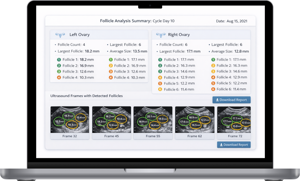
We implemented Mask R-CNN (using ResNet) and extended it with cross-frame tracking, so the system follows the same follicle across multiple frames. Pixel masks are into meaningful units using a calibration module that detects the ultrasound hatch marks (5 mm spacing), computes a pixel-to-mm ratio, and so calculates diameters, perimeter, and surface area per follicle.
Pre- and post-processing removes ultrasound noise and low-contrast boundaries, so a clinician sees only clear results and can track validation metrics. The software supports videos (which are automatically splitted into frames), single images, and folders.
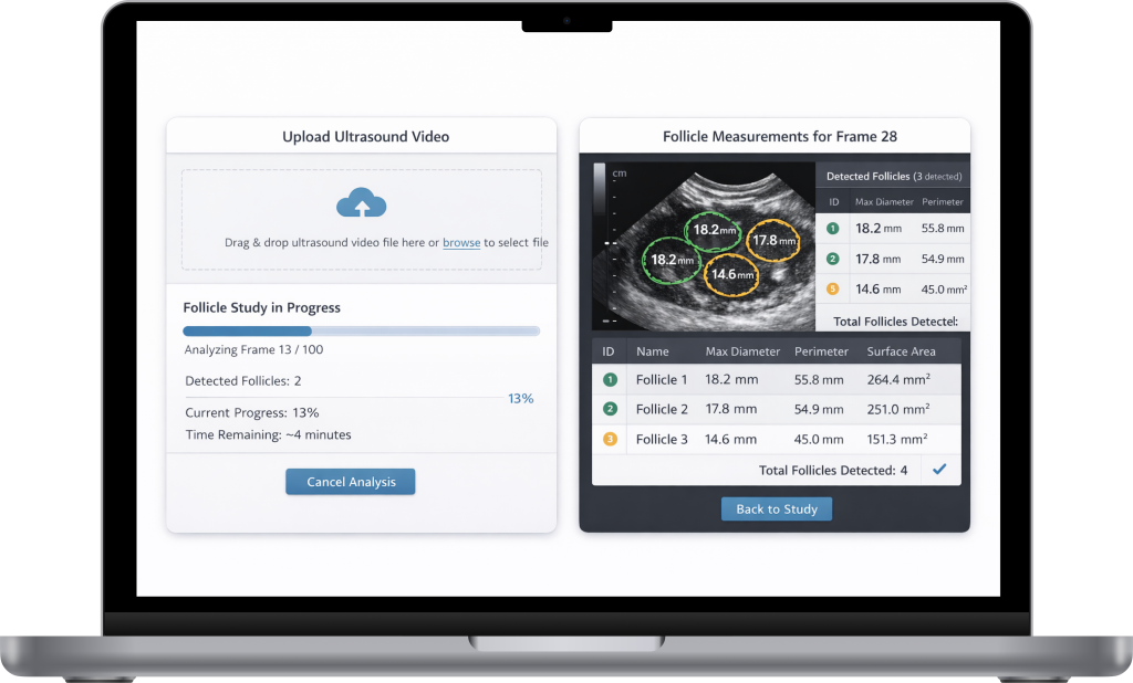
Multi-format ingestion and frame extraction
Accepts ultrasound videos, single images, and folders; automatically extracts frames from video inputs and normalizes them into a consistent processing format.
Ultrasound-specific processing pipeline
Applies filtering and refinement steps tailored to noisy, low-contrast ultrasound data to stabilize contours and reduce artifacts before measurement is computed.
Validation and performance module
Generates precision/recall analytics and run-level summaries to support clinical validation, iterative training, and reproducible QA cycles.
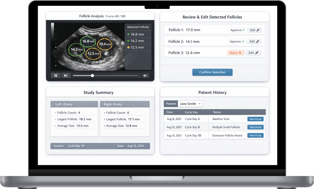
Review-ready output packaging
Exports measured findings in a structured, clinician-friendly format (per study/per frame/per detected instance), enabling quick verification and traceability.
Multi-format ingestion and frame extraction
Accepts ultrasound videos, single images, and folders, automatically extracts frames from video inputs and normalizes them into a consistent processing format.